- Mail At drsantanu.surg@gmail.com
- Call For An Appointment (+91) 9830884263
- Online Chatting (+91) 9830884263
"General Surgery" is a discipline of surgery having a central core of knowledge embracing anatomy, physiology, metabolism, immunology, nutrition, pathology, wound healing, shock and resuscitation, intensive care, and neoplasia, which are common to all surgical specialties.
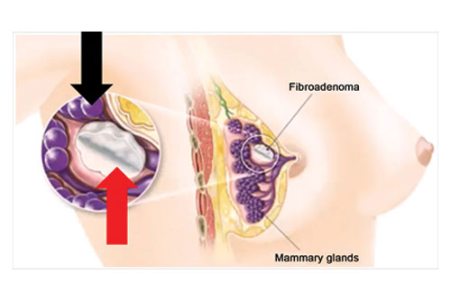
Breast lump removal is surgery to remove a lump that may be breast cancer. Tissue around the lump is also removed. This surgery is called an excisional breast biopsy, or lumpectomy. When a noncancerous tumor such as a fibroadenoma of the breast is removed, this is also called an excisional breast biopsy, or a lumpectomy. Breast lump removal is done as an outpatient surgery most of the time. You will be given general anesthesia (you will be asleep, but pain free) or local anesthesia (you are awake, but sedated and pain free). The procedure takes about 1 hour. The surgeon makes a small cut on your breast. The cancer and some of the normal breast tissue around it is removed. A pathologist examines a sample of the removed tissue to make sure all the cancer has been taken out.
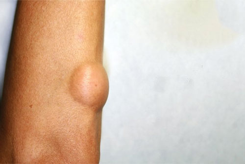
Larger lipomas are best removed through incisions made in the skin overlying the lipoma. The incisions are configured like a fusiform excision following the skin tension lines and are smaller than the underlying tumor. The central island of skin to be excised is grasped with a hemostat, or Allis clamp, which is used to provide traction for the removal of the tumor (Figure 3). Dissection is then performed beneath the subcutaneous fat to the tumor. Any tissue cutting is performed under direct visualization using a no. 15 scalpel or scissors around the lipoma. Care must be taken to avoid nerves or blood vessels that may lie just beneath the tumor.
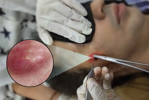
Excision of a sebaceous cyst is a common procedure carried out in plastic and general surgical departments and in general practice. The cyst should be excised completely in order to avoid recurrence. If the cyst ruptures during the procedure there is an increased infection risk and recurrence rates are higher. Several methods have been employed for excising sebaceous cysts
Procedures to remove a fibroadenoma include: 1. Lumpectomy or excisional biopsy. In this procedure, a surgeon removes breast tissue and sends it to a lab to check for cancer. Cryoablation. Doctor inserts a thin, wand-like device (cryoprobe) through your skin to the fibroadenoma. A gas is used to freeze and destroy the tissue. After a fibroadenoma is removed, it's possible for one or more new fibroadenomas to develop. New breast lumps need to be assessed with a mammogram, ultrasound and possibly biopsy — to determine if the lump is a fibroadenoma or might become cancerous.
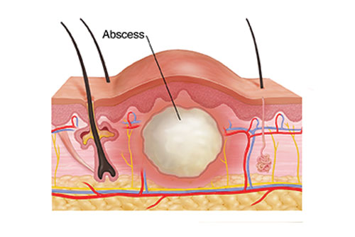
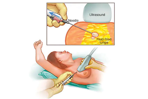
A skin abscess is a pocket of pus just under the surface of an inflamed section of skin. It’s usually triggered by a bacterial infection. Abscess drainage is the treatment typically used to clear a skin abscess of pus and start the healing process. Smaller abscesses may not need to be drained to disappear. Before a skin abscess drainage procedure, you may be started on a course of antibiotic therapy to help treat the infection and prevent associated infection from occurring elsewhere in the body.
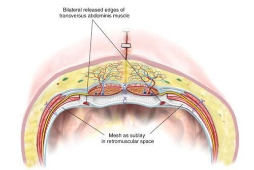
Component separation is an abdominal wall reconstructive technique that strategically divides the rectus and lateral abdominal wall musculofascial layers in order to achieve tension-free midline fascial approximation. Depending on the muscle(s) divided, the techniques of component separation can be broadly categorized into anterior and posterior. Posterior component separation techniques include the Rives-Stoppa retrorectus dissection and transversus abdominis release (TAR).
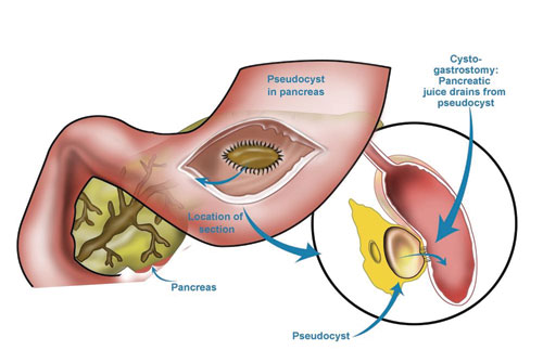
With regard to anatomy, cystogastrostomy was performed for pancreatic pseudocysts directly adherent to the posterior wall of the stomach. Cystoduodenostomy was performed when the cyst was located in the head and uncinate of the pancreas. Roux-en-Y cystojejunostomy was performed for all types of cysts. Surgical resection was used as an alternative approach for pancreatic pseudocysts, and indications for this procedure included cystic neoplasia, splenic vein involvement, upper gastrointestinal bleeding, and technical inability to drain a pseudocyst located in the uncinate.

Surgical staplers and staples are medical devices that may be used in place of sutures. They can close large wounds or incisions more quickly and be less painful than stitches for patients. They are often used in minimally invasive surgery. They can also be used to close wounds in areas where skin is tight against bone, in operations to remove organs or to reconnect parts of internal organs. Surgical staplers are generally made of plastic and loaded with a disposable cartridge of surgical staples. The staplers come in both reusable and disposable models. They resemble construction or industrial staplers and are designed to insert and close several staples at once.
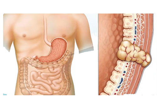
Gastrointestinal stromal tumors (GISTs) are soft-tissue sarcomas that can be located in any part of the digestive system. Their most common sites are the stomach and small intestine. GISTs start in specialized nerve cells located in the walls of your digestive system. These cells are part of the autonomic nervous system. A specific change in the DNA of one of these cells, which control such digestive processes as movement of food through the intestines, gives rise to a GIST. GISTs can develop in people of all ages, but they are most common between age 50 and 70, and they almost never occur before age 40. In rare cases, an inherited genetic change (mutation) causes GISTs.
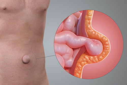
An umbilical hernia occurs when part of your intestine bulges through the opening in your abdominal muscles near your bellybutton (navel). Umbilical hernias are common and typically harmless. Umbilical hernias are most common in infants, but they can affect adults as well. In an infant, an umbilical hernia may be especially evident when the infant cries, causing the bellybutton to protrude. This is a classic sign of an umbilical hernia. Children's umbilical hernias often close on their own in the first two years of life, though some remain open into the fifth year or longer. Umbilical hernias that appear during adulthood are more likely to need surgical repair.
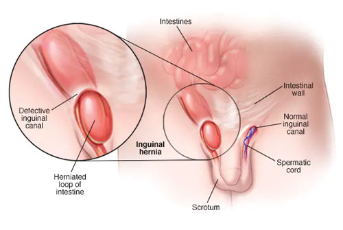
An inguinal hernia occurs when tissue, such as part of the intestine, protrudes through a weak spot in the abdominal muscles. The resulting bulge can be painful, especially when you cough, bend over or lift a heavy object. However, many hernias do not cause pain. An inguinal hernia isn't necessarily dangerous. It doesn't improve on its own, however, and can lead to life-threatening complications. Your doctor is likely to recommend surgery to fix an inguinal hernia that's painful or enlarging. Inguinal hernia repair is a common surgical procedure.
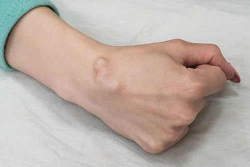
A ganglion cyst is a fluid-filled noncancerous lump that usually develops in the wrist or hand. But some occur in the ankles or feet. When a ganglion cyst presses on a nerve it can be painful. And depending on its location, a ganglion cyst may restrict movement.Some cysts do not need treatment, but others must be surgically removed. During a ganglion cyst removal, a doctor removes the cyst capsule or stalk to completely remove the cyst. Even with surgery, a ganglion cyst may reoccur. Ganglion cyst removal is usually an outpatient procedure and may be performed under local or general anesthesia.
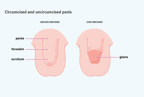
Circumcision is the surgical removal of the skin covering the tip of the penis. The procedure is fairly common for newborn boys in certain parts of the world, including the United States. Circumcision after the newborn period is possible, but it's a more complex procedure. For some families, circumcision is a religious ritual. The procedure can also be a matter of family tradition, personal hygiene or preventive health care. For others, however, circumcision seems unnecessary or disfiguring.
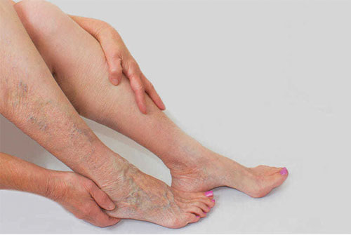
To diagnose varicose veins, a health care provider might recommend a test called a venous Doppler ultrasound of the leg. A Doppler ultrasound is a noninvasive test that uses sound waves to look at blood flow through the valves in the veins. A leg ultrasound can help detect a blood clot. In this test, a health care provider moves a small hand-held device (transducer), which is about the size of a bar of soap, against the skin over the body area being examined. The transducer transmits images of the veins in the legs to a monitor, which displays the results.
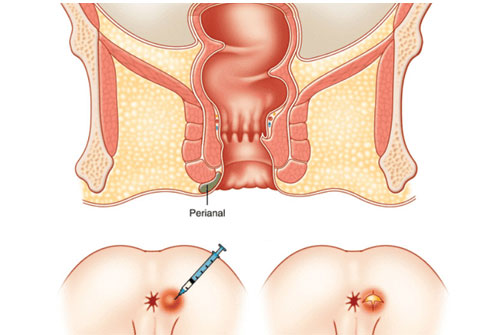
Benign anorectal conditions produce anal pain, rectal bleeding, or discharge from the perianal region, which are highly prevalent symptoms in the general population. Hemorrhoidal disease, anal fissure, perianal abscess, proctalgia syndromes, and pruritus anii are the most common clinical disorders.
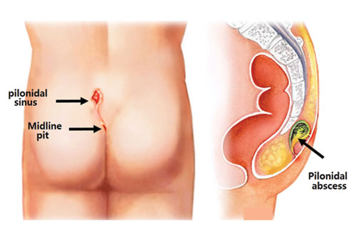
Pilonidal cysts develop near the crease between the buttocks, known as the intergluteal cleft. An impacted or ingrown hair follicle, along with debris like dead skin cells and dirt, become enclosed in a pocket that forms a cyst. The ingrown hair often continues to grow under the skin, irritating the cyst leading to infection. A pilonidal abscess or boil forms and fills with foul-smelling pus. For most patients, the abscess erupts through the skin, draining pus. A pilonidal cystectomy is often required to clear out the infection. Pilonidal cystectomy is a minor surgical procedure that is typically scheduled and performed by a colorectal surgeon on an outpatient basis. General or regional anesthesia may be used to manage pain during the removal of an infected pilonidal cyst or abscess.
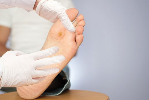
Diabetic foot is a serious complication of diabetes which aggravates the patient’s condition whilst also having significant socioeconomic impact. The aim of the present review is to summarize the causes and pathogenetic mechanisms leading to diabetic foot, and to focus on the management of this important health issue. Increasing physicians’ awareness and hence their ability to identify the “foot at risk,” along with proper foot care, may prevent diabetic foot ulceration and thus reduce the risk of amputation. Diabetic foot ulceration is a major health problem and its management involves a multidisciplinary approach. This review aims to provide a synopsis of the current management strategies of diabetic foot ulcers, from prevention to the options for treatment. The authors believe that it may be useful to primary care physicians, nurses, podiatrists, diabetologists, and vascular surgeons, as well as all healthcare providers involved in the prevention or management of diabetic foot ulcers.
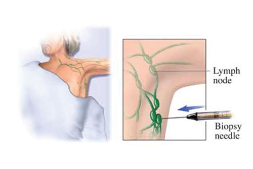
A lymph node biopsy is the removal of lymph node tissue for examination under a microscope. The lymph nodes are small glands that make white blood cells (lymphocytes), which fight infection. Lymph nodes may trap the germs that are causing an infection. Cancer can spread to lymph nodes. A lymph node biopsy is often done in an operating room in a hospital or at an outpatient surgical center. The biopsy may be done in different ways. An open biopsy is surgery to remove all or part of the lymph node. This is usually done if there is a lymph node that can be felt on exam. This can be done with local anesthesia (numbing medicine) injected into the area, or under general anesthesia.
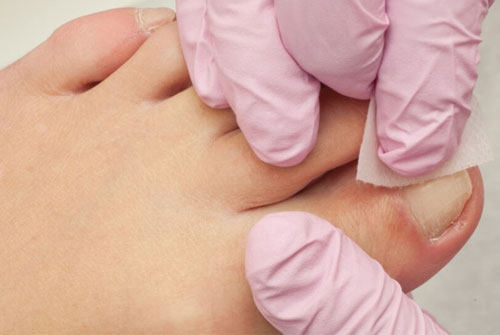
An ingrown toenail is a foot condition that develops when the corner of the toenail grows down into the skin. It usually affects the big toe. Ingrown toenails often happen when people cut their toenails by tapering the corner of their toenail. If the toenail curves with the shape of the toe, it can grow into your skin. Ingrown toenails are common and don’t usually pose a health risk to healthy people. An ingrown toenail can have many causes:
- Incorrectly cut toenails. If you cut your toenails too short or rounded, the nail may grow into the skin.
- Improperly fitting shoes.
- Tearing the corner of the nail.
- Toe trauma, such as banging your toe or getting stepped on.
- Congenital (your foot shape) — for instance, if your nail is larger comparatively with your toe, or the surrounding tissue of the nail border naturally grows around your nail.
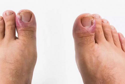
Paronychia (nail infection) usually results from bacteria. Bacteria get into the skin through cuts in the cuticle and the nail fold (the skin around the nail). Most nail infections get better with antibiotics. Paronychia doesn’t usually cause serious health problems. In some cases, the infection lasts a long time or comes back after treatment. Paronychia is nail inflammation that may result from trauma, irritation or infection. It can affect fingernails or toenails. Paronychia can develop when bacteria enter broken skin near the cuticle and nail fold, causing an infection. The cuticle is the skin at the base of the nail. The nail fold is where the skin and nail come together.

Copyright @2021 Dr. Santanu Sarkar. Designed Kolkata Infotech Solution